Department of Surgery, Lagos State University College of Medicine and Lagos State University Teaching Hospital, Ikeja, Lagos, Nigeria
Multiple thyroid ectopia with a normally located thyroid gland is a very rare condition. We present a 34-year old Nigerian woman who had had anterior and left-sided lateral neck swellings for the past 15 years. She was clinically euthyroid and thyroid function test results were normal. Neck ultrasonography revealed multinodular goiter with extension to the left pre-auricular area. Fine Needle Aspiration Cytology (FNAC) was negative for malignancy. She underwent sub-total thyroidectomy and complete excision of three distinct lateral neck masses. Histopathology of the four tissue specimens showed that they were adenomatous goiter without evidence of malignancy. Benign, multiple, laterally located ectopic goiter in the neck can co-exist with normally located multinodular goiter. We report the first case of three ectopic thyroid tissues in the lateral neck co-existing with a normally located multinodular goiter. Although this entity is rare, it should be considered in the differential diagnosis of multiple neck masses.
Ectopic goiter, Multinodular goiter
INTRODUCTION
Thyroid gland development during embryogenesis involves its migration from the foramen caecum at the base of the tongue to its normal pretracheal position. This process can be arrested at any point along the line of descent, resulting in an ectopic thyroid. Although the majority of thyroid ectopias are located in the midline along the tract of the thyroglossal duct, ectopic thyroid tissues can be found in other locations.1
Ectopic thyroid tissue located laterally in the neck were referred to in the past as ‘lateral aberrant thyroid tumours’ because they were thought to represent metastasis from thyroid carcinoma. However, cases of laterally situated benign ectopic thyroid in the neck have been reported.2
We present a rare case of three benign ectopic thyroid masses in the lateral neck region co-existing with a normally located multinodular goiter.
CASE PRESENTATION
A 34-year old female patient presented with anterior and left-sided lateral neck masses first noted 15 years ago. The masses were painless and had been gradually increasing in size over the years. She sought treatment in another hospital ten years ago but declined surgical intervention then offered to her. She had not lost weight and there was no change in her voice. She did not complain of symptoms that could suggest hyperthyroidism, or hypothyroidism.
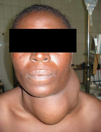
On examination, she had an anteriorly located neck mass of about 13 by 9cm. The mass was firm in consistency, non-tender and moved with deglutition. There were three left-sided lateral neck masses which were firm, non-tender and had smooth surfaces. The largest among them measured 9cm by 7cm, while the smallest measured 3cm by 2.5cm (Figures 1 and 2). 
Figure 1.
Figure 2.
Results of thyroid function test were normal: TSH was 1.1 mU/ml (normal range = 0.54-3.7), T3 was 1.3 ng/ml (normal range = 0.8-2.0) and T4 was 50 ng/ml (normal range = 45-115). Neck ultrasonography revealed an enlarged multinodular left lobe of the thyroid gland with extension laterally to the left pre-auricular area, displacing the left carotid laterally and posteriorly. Fine needle aspiration cytology of the four masses was negative for malignancy.
She was submitted to subtotal thyroidectomy and excision of the three lateral neck swellings through the same collar incision in the neck. The lateral neck masses were separate from each other and from the main gland (Figure 3). Histopathological examination of all specimens revealed features of adenomatous goiter with a section of the lateral masses showing focal papillary formation. There was no evidence of malignancy or lymph node architecture in any specimen. She did well post-operatively and remained clinically and biochemically euthyroid six months after surgery. 
Figure 3.
DISCUSSION
Ectopic thyroid is the most frequent form of thyroid dysgenesis, accounting for 48-61% of the cases.1 Although the incidence of thyroid ectopy is not known, post-mortem studies have suggested that 7-10% of adults may harbour asymptomatic thyroid tissue along the path of the thyroglossal duct.3 Ectopic thyroid tissue co-existing with a eutopic thyroid may be equal to that without a normally located gland.4
Lingual thyroid is the commonest type, while sublingual types are less frequently encountered. The sublingual types could be suprahyoid, infrahyoid or at the level of the hyoid bone.5 Other locations in the neck region where ectopic thyroid tissue may be found include the trachea,6 submandibular7 and lateral cervical regions.1 Furthermore, the presence of ectopic thyroid tissue in other places distant from the neck region has been reported. These sites include the heart,8 duodenum,9 adrenal gland,10 uterus11 and in the Porta Hepatis.12
Although the molecular mechanisms involved in thyroid dysgenesis are not fully known, studies have shown that mutations in regulatory genes expressed in the developing thyroid could be responsible.1 Studies in animals also showed a possible link between development of major cervical arteries and relocalization of the thyroid gland.13
Thyroid ectopia may be associated with thyroid dysfunction, which could be either hypofunction1 or hyperfunction.14 Seventy percent of patients with ectopic lingual thyroid without a co-existing eutopic thyroid tissue will develop sub-clinical hypothyroidism. This often progresses to become clinically manifest during periods of physiological stress.15 Rarely, benign or malignant neoplastic changes can occur in ectopic thyroid tissue.16
Laterally located ectopic thyroid in the neck initially reported, contained malignant tissue and were considered as “aberrant thyroid tumours”. Subsequent reports however described benign ectopic thyroid tissue in the lateral neck region.2,7,17 Ectopic thyroid tissue can present as lateral neck mass without an associated normally located pretracheal thyroid gland. On the other hand, it may co-exist with eutopic thyroid tissue.18 Existence of ectopic thyroid glands at two different locations is very rare. Only 24 cases of such dual ectopia have been reported.19 Huang et al20 reported the second case of dual ectopia with a normally located pretracheal thyroid gland. Presence of three separate ectopic thyroid masses in the lateral neck region with a co-existing eutopic goiter, as seen in our patient, has not been reported in the English literature. It is also unusual that TSH values were not increased in the presence of borderline thyroxine values and such impressive thyroid tissue enlargement.
Though the diagnosis of ectopic thyroid could be difficult, ectopic goiter should be considered in the clinical work-up of neck masses. High resolution ultrasound scanning is useful in the initial assessment, especially in the recognition of a normally located thyroid gland. Diagnosis is usually made by FNAC, while radioisotope scanning using technetium 99m is carried out to identify functioning thyroid tissue. Other imaging studies like computerized tomography (CT) and magnetic resonance imaging (MRI) may be necessary for further evaluation.
Surgical excision of such large ectopic thyroid masses is recommended.
REFERENCES
1. Felice MD, Lauro RD, 2004 Thyroid Development and its disorders: Genetic and molecular mechanisms. Endocrine Reviews 25: 722-746.
2. Stanton A, Allen-Mersh TG, 1984 Is laterally-situated ectopic thyroid tissue always malignant? J R Soc Med 77: 333-334.
3. Sauk JJ, 1970 Ectopic lingual thyroid. J Pathol 102: 239-245.
4. Radkowski D, Arnold J, Healy G, et al, 1991 Thyroglossal duct remnant. Pre-operative evaluation and management. Arch Otolaryngol Head Neck Surg 117: 1378-1381.
5. Batsakis JG, El-Naggar AK, Luna MA, 1996 Thyroid gland ectopias. Am Otol Rhinol Laryngol 105: 996-1000.
6. Hari CK, Brown MJ, Thompson I, 1999 Tall cell variant of papillary carcinoma arising from ectopic thyroid tissue in the trachea. J Laryngol Otol 113: 183-185.
7. Aguirre A, de la Piedro M, Ruiz R, Portilla J, 1991 Ectopic thyroid tissue in the submandibular region. Oral Surg Oral Med Oral Pathol 71: 73-77.
8. Casanova JB, Daly RC, Edwards BS, Tazelaar HD, Thompson GB, 2000 Intracardiac ectopic thyroid. Ann Thorac Surg 70: 1694-1696.
9. Takahasi T, Ishikura H, Kato H, Tarabe T, Yoshiki T, 1991 Ectopic thyroid follicles in the submucosa of the duodenum. Virchows Arch A pathol Anat Histopathol 418: 547-550.
10. Shiraishi T, Imai H, Fukutome K, Watanabe M, Yatani R, 1999 Ectopic thyroid in the adrenal gland. Hum Pathol 30: 105-108.
11. Yilmaz F, Uzunlar AK, Sogutau N, 2005 Ectopic thyroid tissue in the uterus. Acta Obstet Gynaecol Scand 84: 201-202.
12. Ghanem N, Bley Y, Altehoefer C, Högerle S, Langer M, 2003 Ectopic thyroid gland in the Porta Hepatis and Lingua. Thyroid 13: 503-507.
13. Alt B, Elsalini OA, Schrumpf P et al, 2006 Arteries define the position of the thyroid gland during developmental relocalization. Development 136: 3797-3804.
14. Kumar R, Gupta R, Bal CS, Khullar S, Malhotra A, 2000 Thyrotoxicosis in a patient with submandibular thyroid. Thyroid 10: 363-365.
15. Larochelle D, Arcand P, Belzile M, Gagnon NB, 1979 Ectopic thyroid tissue – a review of the literature. J Otolaryngol 8: 523-530.
16. Tucci G, Rulli F, 1999 Follicular Carcinoma in ectopic thyroid gland. A case report. G Chir 20: 97-99.
17. Sahu SK, Agarwal PK, Husain M, Harsh M, Chauhan N, Sachan PK, 2007 Right supraclavicular Ectopic Thyroid: An unusual site of presentation. The internet journal of surgery 13: Number 1.
18. Maino K, Skelton H, Yeager J, Smith KJ, 2004 Benign ectopic thyroid tissue in a cutaneous location: a case report and review. J cutan Pathol 31: 195-198.
19. Chawla M, Kumar R, Malhotra A, 2007 Dual ectopic thyroid: case series and review of the literature. Clin Nucl Med 32: 1-5.
20. Huang TS, Chen HY, 2007 Dual thyroid ectopia with a normally located pretracheal thyroid gland: case report and literature review. Head Neck 29: 885-888.
Address for correspondence:
Ibrahim NA, Department of Surgery, Lagos State University
College of Medicine and Lagos State University Teaching
Hospital, P.M.B. 21266, Ikeja, Lagos, Nigeria
E-mail: ibrahimakanmu@yahoo.com
Received 01-01-09, Revised 15-02-09, Accepted 01-03-09